
Our Office
3125 Coffee Rd.
Suite 1
Modesto, CA 95355
Existing Patients: (209) 529-2726
New Patients: (209) 207-5740
Visit Us Online

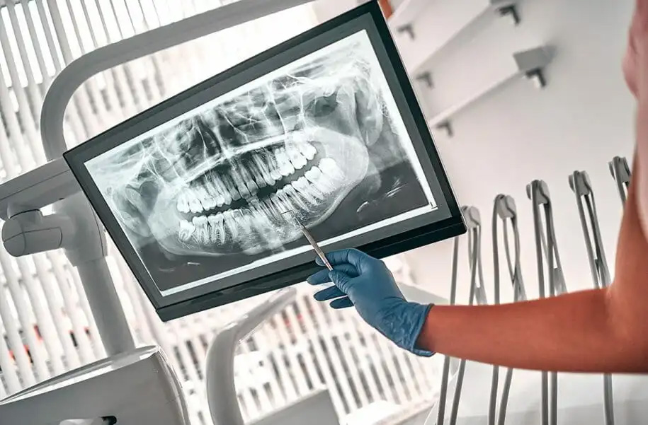
Dental radiographs, commonly known as X-rays, have been an essential tool in dentistry for many years. They provide valuable insights into the internal structures of teeth and the surrounding oral tissues, allowing dentists to make accurate diagnoses and develop appropriate treatment plans. Unlike traditional film-based X-rays, which require chemical processing to produce images, digital radiography offers immediate results that can be viewed on a computer screen. This digital format enables our dentists at Wayne T. Yee, DDS, to enhance and analyze the images, leading to more accurate diagnoses and better patient care.
Digital radiographs provide high-resolution images that allow dentists to identify dental caries in their early stages. Early detection enables prompt intervention and minimally invasive treatments, preserving more of the natural tooth structure.
Digital radiography provides detailed images of teeth, roots, and the surrounding bone, allowing dentists to evaluate the overall health of the dentition and supporting structures.
Digital radiographs help in diagnosing gum diseases and evaluating bone loss around teeth. Periodontal conditions can be monitored over time, enabling dentists to create personalized treatment plans.
Digital radiography plays a crucial role in orthodontic treatment planning. Orthodontists can assess tooth and jaw alignment, identify impacted teeth, and create precise treatment plans for braces or aligners.
Digital radiographs aid in the planning and placement of dental implants. Dentists can determine the optimal position and angle for implant placement, ensuring a successful and long-lasting outcome.
Digital radiography helps diagnose and monitor dental trauma, such as fractures or root fractures, allowing for timely treatment and management.
Digital radiography has revolutionized dental imaging, providing a more efficient, accurate, and patient-friendly way to capture and analyze X-ray images. With reduced radiation exposure, immediate results, and enhanced image quality, digital X-rays have become an invaluable tool in modern dentistry. If you want to experience the benefits of advanced dental care, contact Wayne T. Yee, DDS, at 3125 Coffee Rd # 1, Modesto, CA 95355, or call (209) 529-2726.
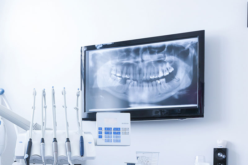
Digital radiography uses electronic sensors to capture images of teeth, roots and surrounding bone instead of exposed film and chemical processing. Images are available instantly on a computer screen, which allows the clinician to review and manipulate the image during the same visit. Because the system is digital, images can be enlarged, contrasted and measured to aid interpretation without degrading the original image.
Compared with traditional film X-rays, digital radiography typically requires less radiation and eliminates chemical waste associated with film development. The immediate availability of images shortens appointment time and supports clearer communication between dentist and patient. Electronic storage also reduces the need for physical space and improves long-term accessibility of records.
Digital radiographs are used to detect a wide range of oral conditions, including dental caries that are not visible on the tooth surface, fractures, infections at the root apex and underlying bone changes. They reveal the shape and length of roots, the presence of impacted or unerupted teeth, and signs of periodontal bone loss. Early detection of these problems supports more conservative and predictable treatment plans.
In addition to caries and trauma, digital images assist in identifying cysts, abnormal growths and changes in bone density that may indicate systemic or localized disease. They are also essential for evaluating the fit and condition of existing restorations and for monitoring healing after procedures. When combined with a clinical exam, radiographs provide a more complete picture of oral health.
Digital radiography uses a fraction of the radiation required by traditional film X-rays because of more sensitive sensors and efficient image capture. Safety protocols such as lead aprons, thyroid collars and modern sensor positioning further minimize exposure to the patient. Dental teams follow the ALARA principle—‘as low as reasonably achievable’—to limit radiation while obtaining clinically useful images.
For most patients, the diagnostic benefits of a properly taken dental radiograph outweigh the minimal risk associated with exposure. Providers determine the timing and frequency of images based on individual risk factors, clinical findings and treatment needs. If you have concerns, your dentist can explain why a particular image is recommended and what steps are taken to reduce exposure.
Digital radiographs provide precise information about tooth anatomy, root morphology and the quality and quantity of alveolar bone, all of which are critical for diagnosis and treatment planning. Dentists use these images to stage periodontal disease, plan endodontic treatment, evaluate for restorative needs and assess the feasibility of implants or extractions. The ability to annotate and measure images supports more accurate procedural planning.
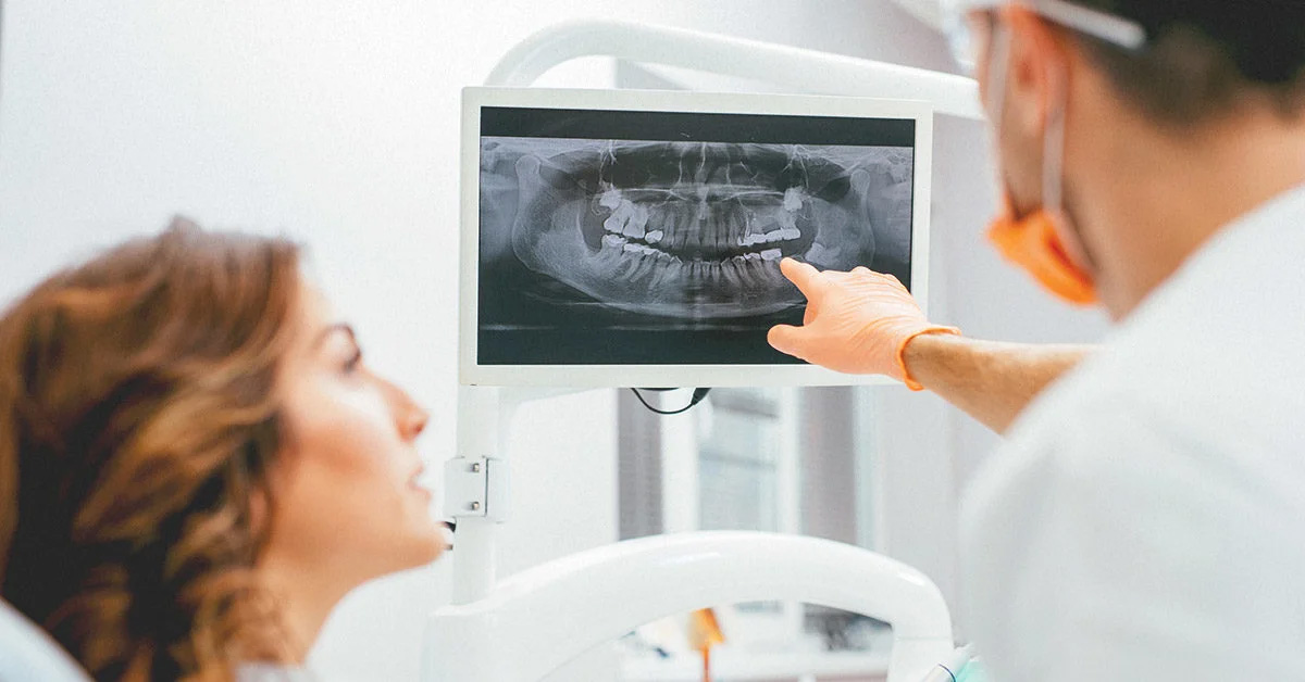
Because images are available immediately, dentists can review findings with patients in real time, discuss options and make timely decisions about care. Serial radiographs can be compared over time to monitor disease progression or healing after treatment. This longitudinal view improves the ability to tailor care to each patient’s changing needs.
High-resolution digital sensors capture fine details that can reveal early-stage decay, small root fractures and subtle bone changes that might be missed on lower-resolution film. Image enhancement tools—such as magnification, contrast adjustment and measurement overlays—help clinicians visualize areas of concern more clearly. These capabilities reduce guesswork and support more definitive diagnoses.
Digital files also allow for side-by-side comparisons, software-aided analysis and integration with other digital records, which enhances clinical decision-making. When multiple specialists are involved, electronic images can be shared quickly to gather additional opinions or plan multidisciplinary care. Overall, digital radiography increases diagnostic consistency and supports evidence-based treatment choices.
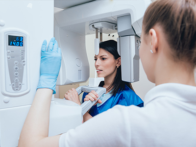
The process is quick and straightforward: a small sensor is positioned inside the mouth while you bite gently to stabilize it, and the image is captured in a matter of seconds. You will be seated comfortably and the dental team will use positioning aids and protective shielding as appropriate. After capture, the image appears on the computer monitor where the dentist reviews it and explains the findings to you.
There is minimal discomfort associated with the procedure, though patients with a strong gag reflex may find intraoral sensors awkward at first; the team can offer techniques to improve comfort during positioning. Because images are ready immediately, any concerns identified can be discussed during the same appointment and incorporated into a treatment plan. At the office of Wayne T. Yee, DDS, the team emphasizes clear communication about what the radiographs reveal and why they are recommended.
Yes, digital radiography systems include tools to adjust brightness, contrast and magnification, and to apply filters that can make specific features more visible. These enhancements do not alter the actual anatomy but help clinicians distinguish between soft tissue, enamel, dentin and bone structures more clearly. Annotating and measuring tools also allow precise assessment of lesion size or root length for procedural planning.
Enhancement features are particularly helpful for identifying early decay, fine root fractures and the extent of bone loss in periodontal disease. Because the original image file is preserved, clinicians can apply different adjustments for teaching, documentation or referral without changing the source image. This flexibility supports careful, repeatable analysis over time.
Digital radiographs are saved as electronic files in a patient’s chart, which enables efficient retrieval, secure backup and long-term recordkeeping without physical film storage. Electronic storage supports HIPAA-compliant security measures such as access controls and encrypted backups to protect patient privacy. When patients transfer care or seek a specialist consultation, images can be exported or shared electronically rather than relying on film copies.
Sharing digital images speeds up referrals and collaborative treatment planning because files can be transmitted quickly and viewed on compatible systems. Many practices also integrate radiographs with practice management and imaging software so clinicians can review images alongside treatment notes and prior records. This integrated approach improves coordination and continuity of care.
Digital radiography can be used safely for children when clinically indicated, with exposure settings adjusted for smaller anatomy and protective shielding in place. Pediatric imaging follows guidelines to limit frequency and scope to what is necessary for diagnosis and treatment planning. Early radiographic assessments may be important for detecting decay between primary teeth, monitoring development and planning orthodontic care.
For pregnant patients, dental radiographs are generally avoided unless essential for diagnosis and treatment that cannot be postponed, and strict shielding protocols are followed when images are required. The dental team evaluates the risks and benefits on a case-by-case basis and uses the lowest practical exposure settings. Open communication with your dentist about pregnancy status allows the team to take appropriate precautions.
Interpreting radiographs requires training, clinical experience and an understanding of how images correlate with intraoral findings; licensed dentists are trained to combine radiographic evidence with a physical exam to arrive at accurate diagnoses. Routine use of digital imaging in clinical practice supports proficiency in reading subtle findings and integrating them into personalized care plans. Modern dental offices also follow evidence-based protocols and quality assurance practices to maintain diagnostic standards.
When complex issues arise, dentists consult with or refer to specialists and may use additional imaging or tests to clarify findings before recommending invasive procedures. If you have questions about an interpretation, ask your dentist to review the image with you, point out specific areas of concern and explain how recommended treatments address those concerns. Wayne T. Yee, DDS and the clinical team prioritize clear explanations so patients understand the rationale behind diagnostic decisions.
