
Our Office
3125 Coffee Rd.
Suite 1
Modesto, CA 95355
Existing Patients: (209) 529-2726
New Patients: (209) 207-5740
Visit Us Online

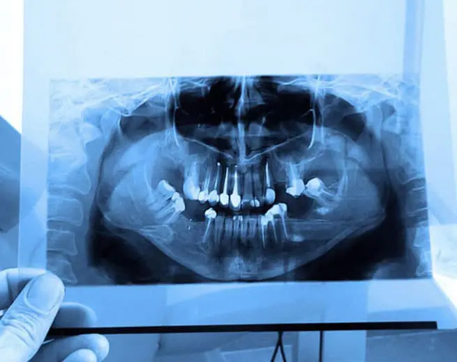
In dentistry, diagnostic imaging is crucial in assessing oral health conditions and planning effective treatments. Panoramic imaging, also known as panoramic radiography or a panoramic X-ray, is a powerful and widely used imaging technique that provides a comprehensive view of the entire oral and maxillofacial region. This non-invasive imaging method offers valuable insights into the teeth, jawbones, sinuses, and other vital structures, aiding dentists and specialists in diagnosing dental issues and developing tailored treatment plans.
At Wayne T. Yee, DDS, panoramic imaging is a specialized dental X-ray technique that captures a two-dimensional, wide-angle image of the entire oral and maxillofacial region. Unlike intraoral X-rays focusing on individual teeth, a panoramic X-ray captures a broader view, showing both the upper and lower jaws, all the teeth, surrounding structures, and the temporomandibular joint (TMJ).
Panoramic imaging is essential to routine dental check-ups and comprehensive dental examinations. It allows dentists to assess the overall dental and skeletal health, identify abnormalities or issues, and create personalized treatment plans.
Panoramic X-rays help dentists evaluate the relationship between teeth, jawbones, and the temporomandibular joint (TMJ). This information is vital for diagnosing malocclusions and planning orthodontic treatments.
Panoramic imaging aids in detecting dental issues such as cavities, impacted teeth, abscesses, cysts, and tumors that may not be visible during a clinical examination.
Panoramic X-rays are beneficial in pediatric dentistry for assessing the growth and development of children's teeth and jaws.
Panoramic imaging is a non-invasive procedure that does not require the placement of X-ray sensors inside the mouth, making it comfortable and suitable for patients of all ages.
Obtaining a panoramic X-ray is quick, usually taking only a few seconds. The immediate results allow dentists to assess the images promptly and discuss the findings with patients.
One of the primary benefits of panoramic imaging is its ability to provide a comprehensive view of the entire oral and maxillofacial region in a single image. This includes the teeth, jaws, temporomandibular joints (TMJ), sinuses, and other facial structures. Dentists can assess multiple areas simultaneously, leading to more accurate diagnoses and treatment planning.
Panoramic X-rays require lower radiation doses than traditional film-based X-rays, making them safer for patients. The reduced radiation exposure is particularly beneficial for individuals needing multiple X-rays or pediatric patients.
The panoramic image offers high diagnostic accuracy, allowing dentists to identify and diagnose various dental issues and oral conditions.
By providing a comprehensive view of the oral cavity, including teeth, jaws, and surrounding structures, panoramic X-rays enable dentists to make informed decisions for better treatment outcomes. For the best dental care tailored to your unique needs, visit Wayne T. Yee, DDS, at 3125 Coffee Rd # 1, Modesto, CA 95355, or call (209) 529-2726.
Panoramic imaging is a diagnostic radiographic technique that produces a single, wide-view two-dimensional image of the entire oral and maxillofacial region. The image captures the upper and lower jaws, all of the teeth, the temporomandibular joints, and portions of the sinuses in one exposure. This noninvasive method gives clinicians a broad overview that supplements clinical examination and more focused intraoral X-rays.
Because it records a large anatomical area in one image, panoramic imaging is especially useful for initial evaluation, screening for large-scale abnormalities, and coordinating care among dental specialists. The technique emphasizes overall relationships and major structural changes rather than extremely fine details. Dentists commonly use panoramic views as part of comprehensive diagnostic workups and treatment planning.
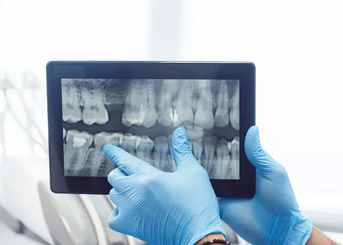
Panoramic imaging captures a wide-angle external view of both dental arches and adjacent structures, while intraoral X-rays focus on individual teeth, tooth roots, and localized areas. Intraoral films and sensor images typically offer higher resolution for detecting small cavities, fine bone detail, and precise root anatomy. Panoramic images are complementary rather than a replacement for intraoral radiographs.
Clinicians often combine panoramic and intraoral images based on the diagnostic task at hand. For example, a panoramic film can screen for impacted teeth or broad bone pathology, and targeted intraoral X-rays can then evaluate suspicious areas at higher resolution. Using both modalities together improves diagnostic confidence and treatment accuracy.
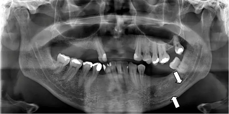
Panoramic X-rays are effective at identifying conditions such as impacted or unerupted teeth, large cysts or tumors, extensive infections, jaw fractures, and gross bone abnormalities. They also reveal the position of wisdom teeth, abnormal jaw growth, and large-scale dental development issues in children and adolescents. These findings can be critical for planning extractions, orthodontic treatment, or oral surgery.
In addition to dental and bony pathologies, panoramic imaging can show sinus changes and some signs of temporomandibular joint involvement, which can guide further evaluation. When a panoramic image suggests a focal lesion or the need for precise bone measurements, the dentist may order more detailed imaging to define the extent and nature of the problem. Overall, the panoramic view is a powerful screening tool for many oral and maxillofacial conditions.
Panoramic imaging exposes patients to a low dose of ionizing radiation, and modern equipment has been designed to minimize exposure while maintaining diagnostic quality. Dental imaging follows the ALARA principle, which stands for "as low as reasonably achievable," so clinicians use the least amount of radiation necessary to obtain useful diagnostic information. Protective measures, such as thyroid collars and careful positioning, further reduce unnecessary exposure.
Certain patient groups, including pregnant patients, require special consideration and discussion with the dentist before imaging is performed. When imaging is clinically indicated, dentists weigh the diagnostic benefits against any potential risks and take appropriate precautions to protect sensitive tissues. If additional reassurance is needed, staff can explain the estimated exposure relative to common background radiation levels.
Preparation for a panoramic X-ray is minimal and typically involves removing metal objects that could interfere with image quality, such as eyeglasses, hearing aids, earrings, necklaces, and hairpins. Patients should also inform the dental team about pregnancy, recent medical imaging, and relevant medical history that could affect imaging decisions. Comfortable clothing and a willingness to remain still during the brief exposure contribute to a clear image.
Children or patients with limited mobility may need brief coaching or assistance from staff to achieve correct head position and stability. There is no fasting or special dietary preparation required prior to panoramic imaging. If a patient has recent dental records or prior images from another provider, bringing them can help the dentist compare changes over time.
During a panoramic appointment the patient stands or sits while the operator positions the head using chin rests, bite blocks, and lateral guides to align the jaws and midline. The machine rotates around the head while a narrow X-ray beam traces the curve of the dental arches, creating the single wide-view image. The actual exposure usually lasts only a few seconds, although total appointment time includes positioning and short image checks by the clinician.
Technicians and dentists review the image promptly to confirm diagnostic quality and to determine whether additional views are needed. The process is noninvasive and generally comfortable, but it relies on patient stillness and correct head alignment for the clearest results. Staff will provide simple instructions and support throughout the procedure.
Panoramic images are interpreted by the treating dentist and, when appropriate, by dental specialists or oral and maxillofacial radiologists for complex findings. The clinicians look for anatomical landmarks, pathology, developmental issues, and relationships between teeth and surrounding bone. If the dentist identifies an area that requires more detailed assessment, they may recommend targeted intraoral films or advanced imaging for clarification.
At the office of Wayne T. Yee, DDS, panoramic radiographs are integrated into comprehensive treatment planning for procedures such as extractions, orthodontics, implant placement, and evaluation of jaw pathology. The images help clinicians communicate findings, explain options, and coordinate care with specialists when multidisciplinary treatment is necessary. Clear radiographic documentation also supports monitoring and follow-up over time.
Panoramic imaging has limitations, including lower resolution for small carious lesions, distortion of structures at the image periphery, and superimposition of anatomical features that can obscure detail. Because of these constraints, panoramic films may not show early-stage cavities or fine root fractures as reliably as intraoral films or three-dimensional studies. Clinicians recognize these strengths and limits when selecting the appropriate imaging modality.
When precise bone measurements, spatial relationships of vital structures, or detailed implant planning are required, a cone beam computed tomography (CBCT) scan or other 3D imaging may be recommended. These advanced scans provide volumetric detail and cross-sectional views that aid in surgical planning and complex diagnoses. Your dentist will recommend 3D imaging only when the expected clinical benefit outweighs additional considerations.
There is no universal interval for panoramic imaging; frequency depends on each patient's clinical history, risk factors, and treatment needs. Dentists individualize imaging schedules based on factors such as age, symptoms, history of disease, orthodontic monitoring, and the presence of implants or other restorative work. Routine use is guided by professional recommendations and the principle of using the minimum imaging necessary for high-quality care.
For example, panoramic images are often obtained as a baseline for new patients, to monitor growth and development in children and adolescents, or to reassess the jaws when there are changes in symptoms. Your dentist will discuss an appropriate imaging plan that balances diagnostic benefit with minimizing exposure to radiation.
To arrange panoramic imaging, call the office at (209) 529-2726 during regular business hours to request an appointment or to discuss any specific concerns about the procedure. Our practice is located at 3125 Coffee Rd. Suite 1 in Modesto, CA 95355, and staff can advise you on the best time and any accommodations you may need for a comfortable visit. When scheduling, please mention if you are a new patient, pregnant, or have mobility needs so the team can prepare appropriately.
When you arrive for your appointment, the dental team will review your medical and dental history, explain the imaging process, and answer any questions before taking the panoramic image. If additional or more detailed imaging is indicated, the dentist will explain the rationale and next steps for diagnosis and treatment planning. Your records and images will be used to develop a safe, effective plan tailored to your oral health needs.
